Calcified hydatid cyst in the liver, seen on CT image (CAT Scan) - stock photo
Benign-looking peripherally calcified cystic lesion within the liver (or spleen) is most in keeping with a hydatid cyst

Get this image in a variety of framing options at Photos.com.
PURCHASE A LICENCE
All Royalty-Free licences include global use rights, comprehensive protection, and simple pricing with volume discounts available
$500.00
+GST NZD
Getty ImagesCalcified Hydatid Cyst In The Liver Seen On Ct Image High-Res Stock Photo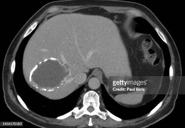 Download premium, authentic Calcified hydatid cyst in the liver, seen on CT image (CAT Scan) stock photos from Getty Images. Explore similar high-resolution stock photos in our expansive visual catalogue.Product #:1456170183
Download premium, authentic Calcified hydatid cyst in the liver, seen on CT image (CAT Scan) stock photos from Getty Images. Explore similar high-resolution stock photos in our expansive visual catalogue.Product #:1456170183$500+GST$50+GST
Getty Images
In stockDETAILS
Credit:
Creative #:
1456170183
Licence type:
Collection:
Moment
Max file size:
8519 x 5869 px (72.13 x 49.69 cm) - 300 dpi - 4 MB
Upload date:
Location:
Romania
Release info:
No release required
Categories:
- Tapeworm,
- CAT Scan,
- Liver - Organ,
- Cancer - Illness,
- Human Digestive System,
- Illness,
- Medical Scan,
- Metastatic Tumour,
- Accuracy,
- Analysing,
- Anatomy,
- Animal Wildlife,
- Biology,
- Black And White,
- Black Background,
- Condition,
- Cyst,
- Data,
- Diaphragm - Body Part,
- Discovery,
- Dog Hookworm,
- Flatworm,
- Healthcare And Medicine,
- Helminthiasis,
- Horizontal,
- Human Body Part,
- Human Internal Organ,
- Human Liver,
- Illustration,
- Indoors,
- Innovation,
- Invertebrate,
- Medical Examination,
- Medical X-ray,
- Medicine,
- Multilocular Adipose Tissue,
- Nature,
- No People,
- Patient,
- Photography,
- Progress,
- Radiologist,
- Research,
- Romania,
- Science,
- Technology,
- The Human Body,
- Tomography,
- Tumour,
- Wound,
- Zoology,